Osteoid Osteoma

Osteoid Osteoma is a benign bone lesion with a nidus of less than 2 cm surrounded by a zone of reactive bone. This lesion accounts for approximately 10 % of benign bone tumors1.The tumor occurs most frequently in the second decade and affects males twice as often as females. The proximal femur is the most common location followed by the tibia, posterior elements of the spine, and the humerus. Osteoid Osteoma is found in the diaphysis or the metaphysis of the proximal end of the bone more often than the distal end.Osteoid osteoma has a distinct clinical picture of dull pain that is worse at night and disappears within 20 to 30 minutes of treatment with non-steroidal anti-inflammatory medication. Joint pain may be present with a periarticular lesion and synovitis can occur secondary to an intraarticular lesion. Local symptoms can include an increase in skin temperature, increased sweating and tenderness. Epiphyseal lesions can cause abnormal growth. The classic radiological presentation of an osteoid osteoma is a radiolucent nidus surrounded by a dramatic reactive sclerosis in the cortex of the bone. The center can range from partially mineralized to osteolytic to entirely calcified. The lesion can occur only in the cortex, in both the cortex and medulla, or only the medulla. The reactive sclerosis may be present or absent. The four diagnostic features include (1) a sharp round or oval lesion that is (2) less than 2 cm in diameter, (3) has a homogeneous dense center and (4) a 1-2 mm peripheral radiolucent zone.' CT is the preferred method of evaluation, especially if the lesion is in the spine or obscured by reactive sclerosis. The radiologic differential includes osteoblastoma, osteomyelitis, arthritis, stress fracture and enostosis.On gross examination, osteoid osteoma is a brownish-red, mottled and gritty lesion that is distinct from the surrounding bone. It can be present in the cortex or medullary canal. Osteoclasts are present. The nidus is surrounded by sclerotic bone with thickened trabeculae.Microscopically, the nidus consists of a combination of osteoid and woven bone surrounded by osteoblasts. The oval shaped nidus is welvascularized and clearly separate from the reactive woven or lamellar bone. Osteoid osteoma will resolve without treatment in an average of 33 months. If the patient does not wish to endure the pain and prolonged use of non-steroidal anti-inflammatory medications, surgical removal or percutaneous ablation of the nucleus is indicated.
References'Bloem, J and H. Kroon, Osseous Lesions, Radiologic Clinics of North America,31(2):261-277, March, 1993. DGitelis, S, Wilkins, R and EU Conrad, benign Bone Tumors, Instructional Course Lectures, 45:425426, 1996.Bullough, Peter, Orthopaedic Patholoev (third edition), Times Mirror International Publishers Limited, London, 1997. Huvos, Andrew, Bone Tumors:Diagnosis, Treatment and Prognosis, W.B. Saunders, Co., 1991.11/18/97


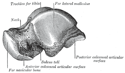
This is a story of my daughter. Truely and frankly, I'm feeling down n upset..terasa mcm ikhtiar yg di buat belum lagi menampakkan hasil. Bile tiap kali terasa perasaan begitu, Istigfar banyak2, yakin: setiap satu penyakit yang Allah turunkan , Allah akan turunkan bersama-sama penawarnya. Cuma kita kene berusaha mencari penawarnya. Penawar ni adalah milik DIA. Jika masih belum lagi ditemukan....mungkin ada hikmah disebaliknya. Cuma terus-terusan seeing her suffering...its a pain to my heart. My fren suggest me to put it in the blog. She said may be ada bloggers have any ideas or suggestion how to help her. She is 17th years old. She was dignosed having ostoeid ostoma late last year after suffering for 5 years. She had to go thru all sort of test since 2004 and until then she was dignosed of having ostoied ostoma. Some says it is a kind non dangerous cancer. It only spread in the bone and will not effect other part of organ in the body. But what happen if the whole bone been effected? Above is picture of the effected bone. Tulang tu tulang sendi kaki kiri. At her ankle. The ostoied ostoma was found at the neck of the bone. The only solution been offered by the doctor is an operation to take-off the effected area. Only that, there is no guarantee that it would be the first and the last operation. The percentage of reoccurance is there. I have meet 3-4 doctors asking for opnions. All said merely the same except that have slight different ideas on how to handle the operation. But the basic thing is:
1. There is no guarantee that the tumor will no reoccure again.
2. When the operation is done, the effected area in the bone will be taken out and will be replace by her bone taken from her hips. Meaning she will have to undergo two operation at the same time. If everything goes well, blood circulation in the bone also goes well, alhamdullillah. Only that, we have to pray that it wouldn't reoccure again.
3. But if the doctor need to scope out the tumor inside the bone in a larger area, there is a posibility of the bone to break. In that case, they have to put screw in order to hold the bone to be stronger. The effected area is at the neck of the bone(middle part). If, they implement the screw, it will disturb the blood flow in the bone and it could end up the bone to be dead. Then they have to take out the whole bone.
She's a girl. My girl. My only girl. I don't know what to do. Seeing her in pain everyday. Hearing her in pain everyday. Sometimes i wish just let me have all her pain.... She always ask me about her condition. What the doctor say. But I don't have the heart to tell her. I don't want she to be depressed when she know about it... I just want her to concentrate on her study, and not to worry about anything... Even i cann't write this story about her in my blog in bahasa coz my tears will always trimbble down like a rain fall...
Mummy really love you and you have to be strong. Coz you are not alone. We always be on your side like a shadow. Always......
Love,
Mummy


Comments